Picture this, doctor: a child comes into your clinic, their mother quite worried, telling you, “Doctor, my child has a strange-looking tooth; it’s like an extra tooth growing from the back.” As soon as you examine them, you confirm her observation—there’s an extra protrusion extending from the cingulum of one of their anterior teeth.
This condition is actually quite well-known and nothing to be alarmed about. Today, we’re going to dive deep into the details of the Talon Cusp, often referred to as “the eagle’s talon.”
What Exactly Is a Talon Cusp?
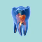
Simply put, a Talon Cusp is a hyperplasia of the cingulum of an anterior tooth, which results in an additional cusp-like structure. So, it’s essentially an extra, small, cusp-shaped piece that grows out from the lingual (inner) surface of the tooth.
What Does It Look Like on X-rays? (Radiographic Features)
On a radiograph, its appearance is quite distinctive, and you can easily spot it if you focus on these key details:
Location
It can appear on any anterior tooth, whether in the primary dentition or the permanent dentition.
Its Edge
The borders of a Talon Cusp are typically very well-defined.
Shape
It generally has a triangular shape. More importantly, it appears superimposed over the crown of the associated tooth on the radiograph itself.
Internal Structure
It’s consistently radiopaque, meaning it appears white on the X-ray, and its radiodensity is similar to that of enamel.
Number
You might find it as a single occurrence, or in some cases, you could encounter multiple Talon Cusps.
Key Points You Absolutely Need to Know
-
It earned its name because of its striking resemblance to an eagle’s talon.
-
The most crucial point, doctor, and one you must underline multiple times, is that it may contain pulp tissue.
-
It’s most commonly found on maxillary anterior teeth, especially the lateral incisors.
Why Is It Clinically Significant? (Clinical Significance)
Despite being a relatively simple condition, a Talon Cusp can sometimes lead to several issues that we need to be mindful of:
-
It can interfere with occlusion, potentially causing problems for the patient when they bite down.
-
Occasionally, its presence may cause aesthetic concerns, particularly if it’s large and noticeable.
-
The surrounding developmental grooves can easily trap food debris, creating a potential for caries development.
-
The most critical aspect: attempting to grind or remove it without meticulous planning could lead to possible pulp exposure. Therefore, extreme caution is absolutely necessary if you decide to intervene.
One Last Note:
Early detection of this condition is vital for proper management. Treatment approaches vary significantly based on its size and the specific problems it’s causing. Options can range from simple monitoring to selective grinding, or even a restorative procedure. All these decisions are made on a case-by-case basis, tailored to each patient’s unique situation.