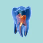
Imagine this, Doctor: you’re taking a routine radiograph for a patient who just came in for a filling, and suddenly, as you’re examining the image, you spot something truly peculiar. You notice that the pulp chamber or root canal in a specific tooth appears anything but normal—it looks as though something is actively eroding and enlarging it from the inside.
This phenomenon has a critically important name in dentistry: Internal Resorption. It’s one of those conditions that, if not diagnosed early, could unfortunately lead to the complete loss of a tooth for your patient.
What Exactly is Internal Resorption?
Simply put, Doctor, internal resorption is a pathological process where the tooth’s structure—be it dentin or enamel—is lost, but crucially, from the inside out. This means the erosion begins within the pulp chamber or root canal and gradually consumes the internal walls of the tooth.
What Does it Look Like on X-Rays? (Radiographic Features)
To accurately diagnose it from radiographs, you absolutely need to pay close attention to these specific details:
Location
It can manifest in any pulp chamber or root canal.
Edge Characteristics
Its borders are typically well-defined and present with a smooth outline. What’s even more important is that these borders are continuous with the existing pulp chamber or root canal; you’ll literally see the canal progressing normally, suddenly widening, and then continuing its usual path.
Shape
The shape can vary from a distinct round appearance to a more linear configuration.
Internal Composition
Internally, it appears radiolucent (transparent to X-rays) because, quite simply, the hard tissue in that area has been resorbed.
Number of Lesions
It can occur as a single lesion or as multiple lesions within the same tooth or in different teeth.
Key Diagnostic Signs
To avoid any confusion, these two critical signs will definitively confirm your diagnosis:
-
You’ll observe an enlarged pulp chamber and/or root canal.
-
You’ll see a distinct radiolucent area within the tooth’s structure itself.
Why is This Clinically Significant? (Clinical Importance)
This condition, Doctor, is far from benign and demands prompt intervention because it can lead to:
-
Significant weakening of the tooth, potentially resulting in fracture.
-
If left untreated, the resorption can progress to the external tooth surface.
-
It is often asymptomatic, which critically emphasizes the importance of routine radiographs for early detection.
-
Once diagnosed, it absolutely requires prompt endodontic intervention to halt this destructive process.
A Crucial Diagnostic Point
Be extremely careful not to confuse internal resorption with external resorption. Differentiating between the two is vitally important. Internal resorption consistently maintains its smooth, well-defined borders that are continuous with the pulp space. In contrast, external resorption typically presents with irregular margins.