Encountering a case of Cleft Palate (or what’s colloquially known as “harelip”) is often one of the most striking and, frankly, daunting situations for anyone not specialized in this field. The moment we see a child with this condition, it’s natural to feel that it’s a vast and incredibly complicated issue. And truthfully, it really does demand substantial effort and a full, dedicated team of medical professionals for proper management.
However, our crucial role as dental professionals, particularly within the radiology department, is to thoroughly understand how the various dimensions of this condition manifest on imaging. This understanding is key to making accurate diagnoses and developing effective treatment plans.
What Exactly is a Cleft Palate? (Defining this Orofacial Anomaly)
Essentially, a cleft palate is considered a congenital defect. It occurs when the maxillary developmental processes fail to fuse correctly during the intricate stages of embryonic formation. To put it more simply, the parts that are supposed to form the roof of the mouth and the lip typically join together at a specific point during development; if this fusion doesn’t happen, a cleft forms.
What Does it Look Like on X-Rays? (Radiographic Features)
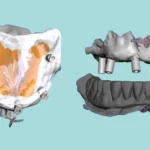
On radiographic images, particularly panoramic views and CBCT scans, we can observe very distinct signs.
Location
-
It is consistently found in the anterior maxilla (the front part of the upper jaw).
Margins
-
The cleft itself will present with well-defined edges, clearly delineated.
Shape
-
Typically, it takes on a linear, line-like configuration.
Internal Structure
-
Radiographically, it appears radiolucent. This is because it’s essentially a void or composed of soft tissue, not bone.
Number
-
It can be a single cleft, occurring on just one side (unilateral).
-
Alternatively, there might be multiple clefts, affecting both sides (bilateral).
Key Diagnostic Signs (Especially for Panoramic Radiographs)
To easily identify and diagnose a cleft palate on a panoramic X-ray, you’ll want to look for these specific indicators:
-
You’ll observe a clear discontinuity of the nasal cavity floor.
-
There will be a distinct vertical radiolucent area, extending downwards from the floor of the nasal cavity.
-
It’s quite common to also find a defect or a cleft within the alveolar ridge itself.
What’s Its Clinical Significance? (Beyond Aesthetics)
This condition isn’t merely about aesthetic appearance; a cleft palate carries substantial clinical implications:
-
Infants with cleft palates often experience significant feeding difficulties.
-
There’s a high likelihood it can lead to speech and hearing problems as the child develops.
-
Quite frequently, it’s associated with various dental anomalies, such as missing teeth or, conversely, supernumerary teeth.
-
Its management invariably requires a multidisciplinary treatment approach, involving a team that might include an orthodontist, oral surgeon, speech therapist, and pediatric dentist, among others.
An Important Final Thought
It’s crucial for us to remember, doctor, that while radiographic examination is an absolutely vital component of both treatment planning and follow-up, the primary diagnosis of this condition remains primarily clinical. Three-dimensional imaging techniques, such as CBCT, offer us incredibly precise details regarding the cleft’s exact size and extent. This information is tremendously beneficial for comprehensive surgical planning.