Imagine this scenario, Doctor. A child or a young adult, typically under 20 years old, walks into your clinic. Their parents are quite concerned because their permanent first mandibular molar seems to be erupting oddly, or perhaps there’s a noticeable swelling on the buccal (cheek) side. Your first logical steps would be a thorough clinical examination and, naturally, some radiographs.
During your examination, you find the molar itself appears perfectly sound—no caries. However, the surrounding gingiva might show some mild inflammation. A vitality test confirms it’s vital. Then, you glance at the radiographs and spot a distinct radiolucency right in the furcation region. This is precisely where you need to consider a very specific and important diagnosis: the Buccal Bifurcation Cyst.
What Exactly Is a Buccal Bifurcation Cyst?
To put it very simply, Doctor, this is an inflammatory cyst, which isn’t exceedingly common. It’s almost always associated with mandibular molars, particularly in younger individuals—those typically under 20 years of age.
How It Appears on X-rays: Radiographic Features
To easily identify it on radiographs, pay close attention to these details:
Location
It’s consistently found in the furcation region of mandibular molars. The most common site is the permanent first mandibular molar, though it’s less frequently seen with second and third molars.
Edge
Its borders are always well-defined and might even be corticated, appearing as a distinct white line.
Shape
It generally presents as round or ovoid.
Internal Composition
Internally, it appears radiolucent, meaning it’s transparent to X-rays, and it’s always unilocular (single-chambered).
Number
It can be a single lesion, but it’s also possible to encounter multiple cysts.
Key Diagnostic Signs
There are a few really distinctive signs you absolutely need to look out for to make the correct diagnosis:
-
The presence of a radiolucent lesion within the furcation region of the molar.
-
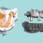
The most critical point: The tooth associated with this cyst is usually tilted lingually, meaning its roots are displaced towards the tongue.
-
As a direct consequence of this lingual tilt, when you perform your clinical examination, you’ll often notice that the lingual cusps appear higher than the buccal cusps.
Clinical Significance
The presence of this cyst can potentially cause tooth displacement or lead to delayed eruption. It might also result in an expansion of the buccal cortical plate. A notable characteristic is that the associated teeth typically remain vital.
Treatment most often involves surgical enucleation followed by careful monitoring.
Important Points to Keep in Mind
-
This cyst goes by a few other names, such as paradental cyst or mandibular infected buccal cyst.
-
Utilizing CBCT imaging can provide a significantly clearer view of the tooth’s angulation and the full extent of the lesion.
It’s absolutely vital, Doctor, to differentiate this from other lesions that can appear in the same area, like periapical or periodontal lesions. The correct diagnosis is truly the cornerstone of appropriate case management.