Imagine this, doctor: you’re examining a patient, and as you check their premolars, you suddenly notice a small, unusual projection on the occlusal surface—almost like a tiny mountain peak rising from the middle of the tooth. What’s the very first thing that comes to mind? Is it decay? Or perhaps a strangely shaped old filling?
The truth is, this is a condition we don’t encounter every day, and it’s called Dens Evaginatus. It’s really important to recognize it and understand how to manage it, because ignoring it can lead to significant problems down the line.
What Exactly is Dens Evaginatus?
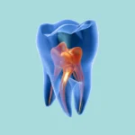
Simply put, doctor, Dens Evaginatus is a developmental anomaly. In this condition, an extra part of the enamel protrudes outwards from either the occlusal or incisal surface of a tooth. Think of it as an additional, small tubercle or projection made of enamel.
Radiographic Features: How It Appears on X-rays
Identifying Dens Evaginatus quickly on radiographs is quite straightforward. Here’s what to look for:
Location
It most commonly appears on lateral incisors and premolars.
Edge Definition
Its borders are typically very clear and well-defined.
Shape
On an X-ray, it usually presents as a distinctive triangular projection.
Internal Structure
It will appear radiopaque, meaning it’s opaque to X-rays, and its density will be similar to that of enamel—an enamel-like density.
Number
Usually, you’ll find just one such projection per tooth, though it’s certainly possible to observe multiple affected teeth in the same patient.
Key Points Every Dentist Should Know
-
This condition is considered less common than its counterpart, Dens Invaginatus.
-
Here’s the most critical and concerning detail, doctor: this projection can often contain a small pulp horn extension. This particular feature is the root cause of nearly all potential problems.
-
As mentioned, you might also find it affecting more than one tooth in a single individual.
Clinical Significance: Why It Matters So Much
Even though it might look unassuming, Dens Evaginatus can unfortunately lead to major complications if not managed correctly:
-
Risk of Pulp Exposure: Given that this projection can house a part of the pulp, any fracture—or even natural attrition over time—can very easily lead to pulp exposure.
-
Occlusal Interference: Occasionally, these projections can interfere with a patient’s normal occlusion.
-
Periodontal Issues: If the projection is situated too close to the gingival margin, it could potentially contribute to periodontal problems.
That’s why early detection is incredibly important. It allows you to take preventive measures and effectively protect the tooth.
So, How Do We Manage It?
Regular monitoring is truly the cornerstone of preventing any complications. The specific treatment approach will depend on the size and location of the projection, and it might involve one of the following:
-
Selective Grinding: This involves very careful and gradual reduction of the cusp over several appointments. This allows the pulp adequate time to form reparative dentin.
-
Bonding: Applying a restorative material to reinforce the projection, thereby protecting it from fracture.
-
Or any other preventive procedures you deem appropriate for the specific case.