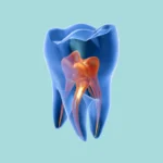
Imagine a young patient walking into your clinic, their smile completely compromised. Their teeth look unusual, discolored in shades of yellow or brown, and appear worn down and fragile. This isn’t just a typical case of neglect; it could very well be Amelogenesis Imperfecta (AI). This condition truly puts a dentist’s skill to the test because it’s far more than just an aesthetic issue—it’s often a significant psychological and social challenge for the patient.
Amelogenesis Imperfecta refers to a group of hereditary disorders that disrupt the proper formation of the enamel layer, leading to weak, easily fractured teeth with an abnormal appearance. Effective treatment requires a multidisciplinary team to fully restore the patient’s smile and self-confidence. In this comprehensive guide, we’ll break down everything you need to know to manage these cases with professional expertise.
What Exactly is Amelogenesis Imperfecta?
Simply put, Amelogenesis Imperfecta (AI) encompasses a diverse group of inherited conditions that disrupt the intricate process of enamel formation—that vital outer layer protecting our teeth. The root of the problem lies in mutations occurring within the genes responsible for building enamel. This genetic issue can be inherited through several patterns:
-
Autosomal Recessive inheritance
-
Autosomal Dominant inheritance
-
X-Linked inheritance
For instance, in the United States, the prevalence of this condition is estimated to be approximately 1 case per 14,000 individuals.
Types of Amelogenesis Imperfecta
We can broadly categorize AI into three primary types, based on the observable phenotype (the visible characteristics) of the teeth:
-
Hypomaturation: In this type, the quantity of enamel formed is normal, but its quality is compromised; it ends up being too soft and excessively porous.
-
Hypomineralized: Here, the enamel quantity is again normal, but it’s remarkably weak, fracturing even from minimal forces.
-
Mixed AI: This presentation is, as the name suggests, a combination of the two aforementioned types.
It’s really important to note that classifying these types can sometimes be a bit tricky because the appearance of the teeth can change significantly after eruption due to everyday friction and wear.
Why Is This Condition So Crucially Important?
The aesthetic appearance of teeth plays an enormous role in a patient’s psychological well-being, especially for adolescents and young adults. A study conducted on teenagers and young adults with AI revealed that they often experience embarrassment and shame regarding their dental appearance, fearing judgment from others. Therefore, when we improve their smile, we are fundamentally enhancing their overall quality of life.
Multidisciplinary Management
Treating a condition like Amelogenesis Imperfecta absolutely demands a fully integrated team of specialized dentists and exceptional communication both among them and with the patient. The comprehensive treatment plan might involve several dental specialties:
-
Restorative Treatment
-
Periodontal Treatment
-
Orthodontic Treatment
-
Oral Surgery
Effective communication within the team and with the patient is the absolute key to treatment success. These complex cases might necessitate extended periods of provisionalization and meticulous coordination among various clinicians and the dental laboratory. Furthermore, the symbiotic relationship between Prosthetic Dentistry and Periodontics is fundamentally essential to ensure the long-term longevity of fixed restorations. This includes designing restorations that empower the patient to maintain excellent oral hygiene and preserve healthy gingival tissues.
Case Report
Here, we’ll present a case involving a young female patient diagnosed with AI. The overarching goal of her treatment was to significantly improve the aesthetic appearance of her teeth, protect the underlying dentin, and minimize the future risk of tooth loss.
-
Patient: A 17-year-old non-smoking female.
-
Chief Complaint: The primary issue was the functional and aesthetic full-mouth reconstruction.
Examination and Diagnosis
The initial comprehensive examination included:
-
Full-Mouth Series of Radiographs
-
Cone-Beam Computed Tomography (CBCT)
-
Intra- and Extra-Oral Photographs
-
Mounted Models
The patient had missing teeth numbers 1, 16, 17, and 32. Tooth number 21 was impacted. Teeth numbers 2, 12, 13, 14, and 18 had existing stainless-steel crowns. Additionally, teeth numbers 6, 7, 8, 9, 10, 20, 22, 23, 24, 25, 26, and 27 had bonded composite restorations.

Dentin was exposed on the clinical crowns of the unrestored teeth, and these teeth exhibited clear signs of occlusal wear or fractures. Many teeth appeared structurally weak and presented with minor carious lesions. Teeth 12 through 14 were super-erupted, tooth 8 was mesially inclined, and tooth 20 was intruded.
The overjet was approximately 2 mm, and the overbite was about 6 mm. The maxillary midline was deviated approximately 1 mm to the right relative to the mandibular midline. Periodontal probing depths ranged from 2 to 4 mm. Despite low plaque levels, the gingiva surrounding the maxillary teeth was notably inflamed.
Radiographs confirmed normal periodontal bone levels and adequate root lengths, with no evidence of endodontic pathology. Furthermore, no tooth mobility was observed.
After thoroughly discussing the findings, treatment options, and potential risks with the patient and her parents, we collectively agreed on a treatment approach involving full-mouth single-unit restorations. Clinical Crown Lengthening procedures and interim provisional crowns were deemed essential to achieve aesthetically pleasing final restorations and maintain a healthy periodontium.
Treatment Plan
All examination results were thoroughly presented to the patient and her mother. We developed an initial digital design and diagnostic wax-up models, which were then reviewed and approved by the patient and her family. The proposed restorative treatment plan included the extraction of teeth numbers 2, 15, 18, 21, and 31.

The case was mounted on an articulator in centric relation, utilizing a facebow and bilateral manual manipulation for model mounting. The centric relation mounting was confirmed intraorally after initial mounting. We decided to increase the patient’s anterior occlusal vertical dimension by 2 mm, which would allow us to achieve an ideal occlusal plane and sufficient thickness for the planned occlusal ceramics.
The final restorations were designed as single-unit tooth-supported crowns. Clinical Crown Lengthening procedures provided sufficient clinical crowns for stable restorations with optimal aesthetics. Following approval of the treatment plan, the patient was referred to an oral surgeon for the extraction of teeth numbers 2, 15, 18, 21, and 31. Once these extraction sites had healed, the patient commenced the restorative phase of treatment.
-
Removal of Existing Restorations and Caries: The first step involved the careful removal of all current restorations and any carious tooth structure.
-
Tooth Preparation and Provisional Placement: Teeth were prepared for crowns, and provisional restorations were placed. PMMA provisional restorations were fabricated using impressions taken from the diagnostic wax-up. Each tooth was thoroughly assessed for restorability, confirming that no endodontic therapy was required.
-
Impressions and Crown Lengthening Assessment: VPS impressions of the crown preparations were taken to create a second digital design and diagnostic wax-up. This information was then used to determine the necessity for Clinical Crown Lengthening Surgery.
-
Clinical Crown Lengthening Surgery: The patient was referred to a periodontist. Following detailed written instructions, the periodontist performed clinical crown lengthening on both maxillary and mandibular tooth sites. Ostectomies were carefully performed to achieve the desired gingival contours and establish a 3 mm distance between the osseous crest and the margins of the provisional restorations. This precisely positioned the gingival margins in their pre-determined locations.
-
Gingival Closure: Gingival flaps were secured using continuous 4.0 polytetrafluoroethylene sutures. Additionally, a maxillary anterior frenectomy was performed. The wound was closed with 4.0 plain gut sutures. At a second surgical appointment, the mandibular tooth sites were treated using the same methodology.
-
Follow-up and Preparation for Final Restorations: The patient was advised to maintain excellent oral hygiene and to use a fluoride mouthrinse. She was permitted to proceed to the final restorative phase of treatment 12 weeks after the last surgical procedure.
-
Provisional Adjustments and Final Impressions: Twelve weeks post-surgery, the general dentist meticulously adjusted the crown preparations and provisional restorations. Impressions were then taken for a second set of provisional crowns. Final design decisions, such as shade selection, were also made at this time.
-
Placement of Lab-Fabricated Provisional Crowns: Lab-fabricated PMMA provisional crowns were placed approximately three weeks later. The patient and her mother approved the tooth shapes, color, bite (occlusion), aesthetics, and phonetics. The patient wore these provisional crowns for about seven months. Throughout this period, pulp vitality was carefully monitored, and no endodontic treatment was found necessary.
-
Final Adjustments and Definitive Impressions: After approximately seven months, final crown margin adjustments were made, and full-arch impressions were taken using heavy and light body VPS.
-
Design and Placement of Final Crowns: At the dental laboratory, the final restoration design was digitally created and approved by the general dentist. The final restorations consisted of high translucent monolithic zirconium crowns, which were cemented using a resin-modified glass ionomer luting cement. Per the patient’s request, the crowns were fabricated in a B1 shade without any additional shade gradations.
Outcome

The final restorations exhibited an exceptionally natural and aesthetically pleasing appearance. The patient was able to maintain excellent oral hygiene, and her periodontium remained perfectly healthy.
Both the patient and her mother reported significant beneficial effects from the treatment. The patient expressed feeling much more confident and comfortable in social settings, smiling more often, and feeling more attractive. Consequently, she was able to interact more effectively with her peers and develop closer relationships.
Discussion
Several treatment options were thoroughly discussed with the patient and her mother. Among these were dental implant-supported restorations. Given that AI is characterized by defects in the formation of enamel, it might seem logical to assume a concomitant increase in the risk of dental caries. However, this is not necessarily true. A systematic review of the literature found a low dental caries susceptibility among AI patients. This could potentially be attributed to the lack of proximal contacts and the elimination of fissures. Therefore, caries risk was not a compelling reason to replace her teeth with dental implants.
Considering the patient’s young age, the possibility of continuous craniofacial growth was a significant concern. Maxillomandibular changes throughout adolescence can lead to complications such as implant infraocclusion (IO) and interproximal contact loss (ICL). A study evaluating single-implant restorations in posterior and anterior regions three months to eleven years post-placement found that 52.8% experienced interproximal contact loss.
A literature review on the prevalence and contributing factors of contact loss between implants and adjacent teeth revealed that ICL can occur as early as three months after prosthetic treatment. Another study identified IO and ICL as the most common complications in implants placed in adolescents.
A retrospective study evaluated young and mature adults with single implants in the anterior maxilla. All patients demonstrated IO of the implant-supported crowns. A more recent literature review concluded that facial skeletal growth and teeth eruption are notable during the second and third decades of life. This study advised that, whenever possible, the placement of an anterior maxillary implant should be delayed in adolescent patients. All these factors collectively diminished enthusiasm for utilizing dental implants in such a young patient.
Effective communication between the patient, dentists, and the dental laboratory is absolutely paramount for treatment success. Precise planning and clear communication become particularly crucial in full-mouth rehabilitation cases. The use of digital planning software significantly aids in facilitating this communication. Patients must have clear and appropriate expectations, and the clinical team and laboratory must commit to delivering on those promises.
Research consistently supports the use of permanent tooth-supported fixed restorations for young AI patients. Material selection in this specific case was primarily guided by the patient’s young age and the critical need for long-term crown success. Zirconia, a crystalline oxide of zirconium, possesses excellent mechanical, optical, and biological properties. Zirconia also exhibits the highest hardness among all restorative materials currently utilized in dentistry. Furthermore, studies consistently demonstrate its nature as a biocompatible biomaterial.
In this particular case, monolithic zirconia crowns were utilized. For the molars, zirconia with a lower yttria content was chosen due to its superior strength. For the remaining teeth, zirconia with a higher yttria content was selected for its enhanced aesthetic value. The choice of a B1 shade was made based on the patient’s specific request.
Clinical Crown Lengthening becomes essential whenever the available clinical crown height is insufficient. The appropriate positioning of the osseous crest and gingival margins ensures restorations that do not violate biologic width and allows for the fabrication of restorations with aesthetically pleasing proportions.
Zirconium-based restorations fabricated using Computer-Aided Design and Computer-Aided Manufacturing (CAD/CAM) technology generally yield superior outcomes in terms of marginal fit, reduction of inflammation, and preservation of periodontal health compared to conventional methods. Moreover, supragingival margins promote better oral hygiene compared to subgingival margins.
A fundamental objective in dentistry is to significantly improve the patient’s quality of life. As previously noted, dental aesthetics profoundly influence the psychological and social well-being of adolescent patients. A systematic review of articles revealed that the majority of studies (88%) reported a correlation between malocclusion or structural dental anomalies and exposure to bullying among young adolescents.
Another study specifically evaluated the Oral Health-Related Quality of Life (OHRQoL) in patients with severe AI both before and after crown treatment. Most of the 26 patients reported substantial improvements in their quality of life following treatment. The patient in this article had a poor self-perception of her smile prior to treatment. Consistent with the research, the patient expressed being “very happy with her smile” after the treatment. Her confidence dramatically improved, allowing her to actively and positively engage within her social environment and with her peers.
Conclusion
Patients afflicted with Amelogenesis Imperfecta can face considerable psychological, social, and dental challenges. Adolescents, in particular, are susceptible to self-perception issues directly related to their oral health. Excellent communication and coordinated care among the dental team and laboratories are absolutely crucial to deliver the most successful possible treatment outcomes for these patients.