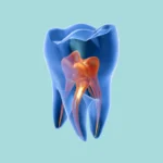
When we talk about Lower Third Molar Impaction, we’re delving into arguably the most common surgical procedure performed in dental clinics. Among the various types, Mesioangular Impaction is the one we most frequently observe on radiographs, characterized by the wisdom tooth tilting forward and impinging on the second molar. The fundamental principle often states that to extract a tooth, you must remove what obstructs its path, which in this case appears to be the mesial portion of the wisdom tooth’s crown. While logic might suggest simply cutting this part to facilitate extraction, the reality is this rule isn’t always correct and can lead to numerous complications, such as root fracture. In this article, we’ll clarify when this decision is appropriate, when it’s not, and how you can define your precise surgical plan just by looking at the X-ray.
Why Sectioning in the First Place? It’s Not Just About “Cutting”
Before we even decide where to cut, we need to ask ourselves a crucial question: Why am I performing sectioning? Sectioning, or dividing the tooth crown, isn’t an end in itself. It’s merely a means to achieve something far more important: creating a clear, unimpeded Path of Withdrawal for the tooth.
Imagine trying to maneuver a large sofa through a narrow doorway. If you attempt to pull it through as is, it’ll get stuck. However, if you detach one of its legs, suddenly you discover new space, allowing you to easily move it out. Sectioning is precisely like detaching that “leg.” We remove a small part of the tooth to open up a smooth pathway, enabling the tooth to exit without fracturing itself or causing damage to adjacent structures.
The Old Rule and Why It’s Not Always Right (Its Inherent Flaw)
The rule many of us were taught often dictates: “The obstruction is mesial, therefore, cut the mesial part.” While this guideline seems perfectly logical at first glance, it crucially overlooks the most significant factor in the entire equation: the Root Curvature.
Wisdom tooth roots are very rarely perfectly straight. More often than not, they are angled either distally (backward) or mesially (forward). Attempting to extract a tooth against the direction of its root curvature is, quite simply, a recipe for disaster. Picture trying to pull a bent nail straight out of a wall; it’s almost guaranteed to break. You must pull it in the same direction as its curve.
The X-ray Holds the Secret: How to Interpret It Correctly (The Key to Your Plan)
Your entire decision-making process happens right at your desk, while you’re examining the radiograph, even before the patient opens their mouth. This is why having a high-quality X-ray (either periapical or panoramic) is essential. You need to meticulously observe three primary aspects, in this specific order:
-
Angulation: Confirm that it is indeed a Mesioangular impaction.
-
Crown Morphology: Assess its size and whether its cusps are prominent and well-defined.
-
The Most Crucial Point: Root Curvature: Pay very close attention to the apical third of the roots. Are they distinctly curved distally (backward)? Are they curved mesially (forward)? Or are they relatively straight?
Based directly on your answer to this third question, you’ll be able to precisely determine your strategic “game plan.”
The Game Plan: When to Go Mesial and When to Go Distal?
Let’s break down each scenario individually and see how we should proceed.
Case 1: Distally Curved Roots (The Most Common Scenario)
This is the situation we encounter in approximately 80% of cases. The roots are clearly curving distally, towards the ramus.
Analysis: Here, the natural path of withdrawal for the tooth must be distally and occlusally. If you try to elevate this tooth mesially, you’re pulling the roots directly against their natural curvature, and the inevitable outcome will be fracture.
Action Plan:
-
The sectioning will be performed on the distal part of the crown. You’ll use your bur to cut the distal cusp and typically a portion of the distal marginal ridge.
-
By doing this, you’ve effectively created space behind the tooth.
-
Next, insert your elevator (such as a Cryer or Crane pick) into the area you just sectioned. Using the second molar (#7) as a fulcrum, gently apply force to dislodge the wisdom tooth distally and inferiorly first (to free it from #7), then distally and superiorly into the void you’ve created.
Result: The tooth will emerge in one piece, following the natural direction of its root curvature, and with minimal effort on your part.
Case 2: Mesially Curved Roots (A Less Frequent Occurrence)
This isn’t a very common scenario, but it certainly happens. Here, the roots are curving mesially, towards the second molar (#7).
Analysis: In this particular case, the natural path of withdrawal for the tooth is mesially and occlusally. Crucially, the old rule (to cut the mesial part) applies 100% here.
Action Plan:
-
The sectioning will be performed on the mesial part of the crown. You’ll use your bur to cut the anterior portion of the crown that is impacted against #7.
-
By doing this, you’ve effectively removed the direct obstruction preventing the tooth from moving.
-
Then, insert your elevator from the buccal aspect and elevate the tooth superiorly and mesially, following the direction of its root curvature.
Result: The tooth will extract easily because you’re working in harmony with the roots’ natural direction.
Case 3: Straight Roots
Analysis: In this situation, there is no inherent directional pull from the roots. You have greater freedom of movement.
Action Plan:
-
Here, the simplest and most logical choice is to revert to the old rule: section the mesial cusp, as it represents the direct mechanical obstruction.
-
After removing this section, you can then elevate the tooth relatively straight upwards or with a slight distal tilt.
Practical Tips for Successful Sectioning
-
Use the Right Bur: The ideal choice is a long shank surgical bur, such as an H.702L or H.703L, to ensure easy access to the surgical site.
-
Don’t Cut Through the Entire Tooth: Only cut about two-thirds to three-quarters of the tooth’s thickness. Then, use an elevator to “fracture” the remaining portion. This vital technique protects you from potentially injuring the lingual nerve, which is often in very close proximity on that side.
-
Continuous Irrigation: Always use a generous amount of sterile saline while cutting. This continuous cooling prevents overheating of the bone, which can otherwise lead to complications during healing.
-
A Solid Fulcrum: Absolutely ensure that your elevator is firmly supported on a strong and stable fulcrum before you begin applying any lifting force.
Conclusion: Think Like a Chess Player
Surgical extraction of a wisdom tooth is far more than just applying brute force; it’s a calculated process of planning and strategic thinking. Before you even touch the patient, carefully analyze the radiograph and thoroughly assess the situation. Never commit to a single rule and apply it indiscriminately to every case.
Always allow the tooth’s roots to dictate the direction you should take. When you respect the tooth’s unique anatomy and work with it rather than against it, you’ll discover that even the most challenging cases resolve with remarkable ease. More importantly, you’ll effectively avoid that dreaded nightmare of root fracture, which is a concern for all of us.