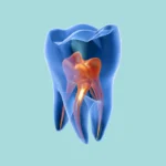
Working with composite resin in posterior teeth can feel like one of those tasks that are deceptively simple yet incredibly challenging. We all perform these restorations almost daily, but the real difference between a filling that “just gets by” and one that truly endures for years without issues lies in the intricate details. These small nuances are precisely what separates a dentist who frequently encounters post-operative sensitivity or restoration fractures from one whose work remains consistently stable and perfectly sealed.
This article isn’t about dry academic textbook theories. Instead, it’s the distilled essence of practical, hands-on work—the concentrated wisdom you need to proactively avoid the most common pitfalls. We’ve meticulously gathered 10 golden secrets, or rather, pivotal guidelines. If you integrate these into your practice, your work will undoubtedly elevate to an entirely new echelon.
1. Isolation Done Right… at the Perfect Moment (Timing is Key)
The paramount rule for composite restorations is complete isolation. However, even more crucial than the isolation itself is its precise timing. A frequent oversight many practitioners make is to begin caries removal before even considering isolation.
The Correct Approach: The rubber dam absolutely must be placed immediately after administering the local anesthetic, and critically, before any bur touches the tooth. Simply put, from the very first moment of excavation, you are exposing the operative field to contamination from saliva and bacteria. By isolating from the outset, you guarantee a pristine, impeccably dry working environment. This is the foundational and most vital prerequisite for successful bonding.
Dr. LOD Tip: When performing a Class II restoration, once the dam is securely positioned, use a wooden wedge. Gently insert it to stabilize the dam sheet in the interproximal area. This protective measure prevents the dam from tearing, especially when you’re operating in deeper regions.
2. Cavity Preparation: Smooth Surfaces are Non-Negotiable (Smooth Cavity Preparation)
Have you removed all the decay? Excellent. But don’t hastily proceed to etching. The surface left by the bur is typically coated with a layer known as the Smear Layer. This layer, composed of dentinal debris and bacteria, unfortunately hinders the optimal adhesion of the bonding agent.
The Correct Approach: After completing the debridement, carefully use a fine diamond stone. Gently glide it over all the cavity walls and margins. This specific action effectively removes the smear layer, preparing a clean tooth surface that is optimally receptive to the bonding agent.
Important Warning: Before executing this step, if you’ve placed a wedge, remove it meticulously and gently. Any gingival bleeding will compromise the entire field. Should bleeding occur, do not continue. Completely staunch the bleeding, then thoroughly clean the area, perhaps with glycerin or a 2% chlorhexidine solution.
3. Smart Matrix Strategy: Think Ahead (A Proactive Approach)
When faced with an MOD cavity, how do you typically strategize? Do you build one proximal wall, then the other?
The Smarter, Faster Way: The most intelligent and efficient method is to place all sectional matrices simultaneously from the outset. While this might add a few extra seconds to your initial setup, it definitively prevents contamination of the cavity and ultimately saves you significant time in the long run.
4. Bonding Technique: The Best of Both Worlds (A Safer, Stronger Bond)
Aspiring for the strongest possible bond with minimized post-operative sensitivity? Contemporary recommendations advocate for integrating two distinct approaches.
The Optimal Method: Implement the Selective Etching technique.
-
Apply phosphoric acid etch only to the enamel, for approximately 15 seconds.
-
Rinse and thoroughly dry the area.
-
Subsequently, apply a 2-Step Self-Etch Bonding Agent to both the dentin and enamel.
This approach harnesses the superior bond strength achieved on enamel while intelligently safeguarding the dentin from excessive etching, which could potentially induce sensitivity.
5. Simplify Your Work: Turning Class II into Class I
Never approach a Class II cavity as a monolithic, unmanageable entity. Instead, segment it to gain better control.
The Strategic First Step: The initial crucial step is to meticulously build up the deficient proximal wall. Construct this wall with composite until it seamlessly matches the level of the marginal ridge of the adjacent tooth. Congratulations! You’ve successfully transformed a challenging Class II cavity into a more manageable Class I.
6. Secure That Wall Before Matrix Removal (Preemptive Reinforcement)
You’ve successfully built the proximal wall. Excellent. While some clinicians might prefer removing the matrix at this point for enhanced visibility, this carries a degree of risk.
The Safer Practice: Ideally, refrain from removing the matrix entirely. However, if absolutely necessary, employ a small reinforcing maneuver: place a thin layer (approximately 1mm) of flowable composite. Use this to create a seamless connection between your newly constructed wall and the internal dentin wall. This clever addition provides crucial structural support, safeguarding it from potential fracture.
7. Your Primary Adversary: Bleeding (Zero Tolerance)
That wedge you’ve carefully positioned isn’t solely for achieving an ideal contact point; it also serves as a critical barrier against bleeding.
The Golden Rule: The cardinal principle is this: do not remove the wedge until you have fully completed every procedural step. Premature removal risks introducing hemorrhage, which can entirely compromise your meticulous work.
8. The Flowable Liner: A Flexible Foundation (Stress Absorption)
Before you commence building the cusps, establish a strategic foundational layer.
The Smart Application: Place a thin layer, approximately 1 mm thick, of flowable composite as a liner on the pulpal floor of the cavity. This layer acts as a “stress absorber,” effectively mitigating the polymerization shrinkage stress of the bulk composite. This significantly reduces post-operative sensitivity.
9. Function Over Flair: Prioritizing Patient Outcomes (Practical Excellence)
We’ve all seen aesthetically stunning composite restorations online. While admirable, artistry isn’t the primary objective.
The True Focus: Concentrate intensely on the elements that genuinely benefit the patient. Your foremost objective is a restoration that:
-
Possesses a precisely adjusted contact point.
-
Is entirely free of any food-trapping overhangs.
-
Exhibits a meticulously balanced occlusion.
-
Features margins that are perfectly sealed.
If you master these critical aspects, you have successfully delivered an exceptional restoration. Intricate sculpting, though desirable, is a refined skill that naturally develops with practice and time.
10. The Finish: Smoothness Ensures Longevity (The Final Polish)
A rough composite restoration inevitably accumulates bacteria and becomes susceptible to staining. Conversely, a highly polished, smooth restoration endures longer and actively contributes to healthier gingival tissues.
The Polishing Protocol: After meticulously completing the restoration and fine-tuning the occlusion, systematically use polishing discs in decreasing abrasive grits. For the definitive final touch, utilize rubber cups or points—particularly the lighter-colored variants such as white or gray—in conjunction with a high-quality polishing paste. This comprehensive finishing regimen will yield a glass-like, lustrous surface, which is the undeniable secret to restorations that truly last for years.